|
Genentech inc
functional blocking antibodies against neuropilin 1 Functional Blocking Antibodies Against Neuropilin 1, supplied by Genentech inc, used in various techniques. Bioz Stars score: 90/100, based on 1 PubMed citations. ZERO BIAS - scores, article reviews, protocol conditions and more https://www.bioz.com/product/rage+function+blocking+antibody/functional+blocking+antibodies+against+neuropilin+1/pmc02741417-89-11-4 Average 90 stars, based on 1 article reviews
functional blocking antibodies against neuropilin 1 - by Bioz Stars,
2026-10
90/100 stars
|
Buy from Supplier |
|
R&D Systems
human s100a9  Human S100a9, supplied by R&D Systems, used in various techniques. Bioz Stars score: 94/100, based on 1 PubMed citations. ZERO BIAS - scores, article reviews, protocol conditions and more https://www.bioz.com/product/rage+function+blocking+antibody/Recombinant+Human+S100A9+Protein%2C+CF/10__2147_slash_ott__s192250-49-5-16 Average 94 stars, based on 1 article reviews
human s100a9 - by Bioz Stars,
2026-10
94/100 stars
|
Buy from Supplier |
|
Alomone Labs
trpv1 epitope specific peptide  Trpv1 Epitope Specific Peptide, supplied by Alomone Labs, used in various techniques. Bioz Stars score: 96/100, based on 1 PubMed citations. ZERO BIAS - scores, article reviews, protocol conditions and more https://www.bioz.com/product/rage+function+blocking+antibody/Human+TRPV1+(extracellular)+Blocking+Peptide/pmc08945112-200-7-10 Average 96 stars, based on 1 article reviews
trpv1 epitope specific peptide - by Bioz Stars,
2026-10
96/100 stars
|
Buy from Supplier |
|
Becton Dickinson
hamster mabs against mouse integrin β3 (2c9.g2  Hamster Mabs Against Mouse Integrin β3 (2c9.G2, supplied by Becton Dickinson, used in various techniques. Bioz Stars score: 90/100, based on 1 PubMed citations. ZERO BIAS - scores, article reviews, protocol conditions and more https://www.bioz.com/product/rage+function+blocking+antibody/anti+%C3%9F3+integrin++2c9+g2/pmc00032268-58-2-17 Average 90 stars, based on 1 article reviews
hamster mabs against mouse integrin β3 (2c9.g2 - by Bioz Stars,
2026-10
90/100 stars
|
Buy from Supplier |
|
Bioss
anti mouse rat human substance p  Anti Mouse Rat Human Substance P, supplied by Bioss, used in various techniques. Bioz Stars score: 90/100, based on 1 PubMed citations. ZERO BIAS - scores, article reviews, protocol conditions and more https://www.bioz.com/product/rage+function+blocking+antibody/Neurokinin+1+Receptor+Antibody%2C+Cy3+Conjugated/pmc07169378-60-0-11 Average 90 stars, based on 1 article reviews
anti mouse rat human substance p - by Bioz Stars,
2026-10
90/100 stars
|
Buy from Supplier |
|
Novus Biologicals
hif1α nb100 134 novus biologicals  Hif1α Nb100 134 Novus Biologicals, supplied by Novus Biologicals, used in various techniques. Bioz Stars score: 95/100, based on 1 PubMed citations. ZERO BIAS - scores, article reviews, protocol conditions and more https://www.bioz.com/product/rage+function+blocking+antibody/HIF-1+alpha+Antibody+-+BSA+Free/10__1071_slash_rd21170-114-26-28 Average 95 stars, based on 1 article reviews
hif1α nb100 134 novus biologicals - by Bioz Stars,
2026-10
95/100 stars
|
Buy from Supplier |
|
R&D Systems
anti goat igg hrp 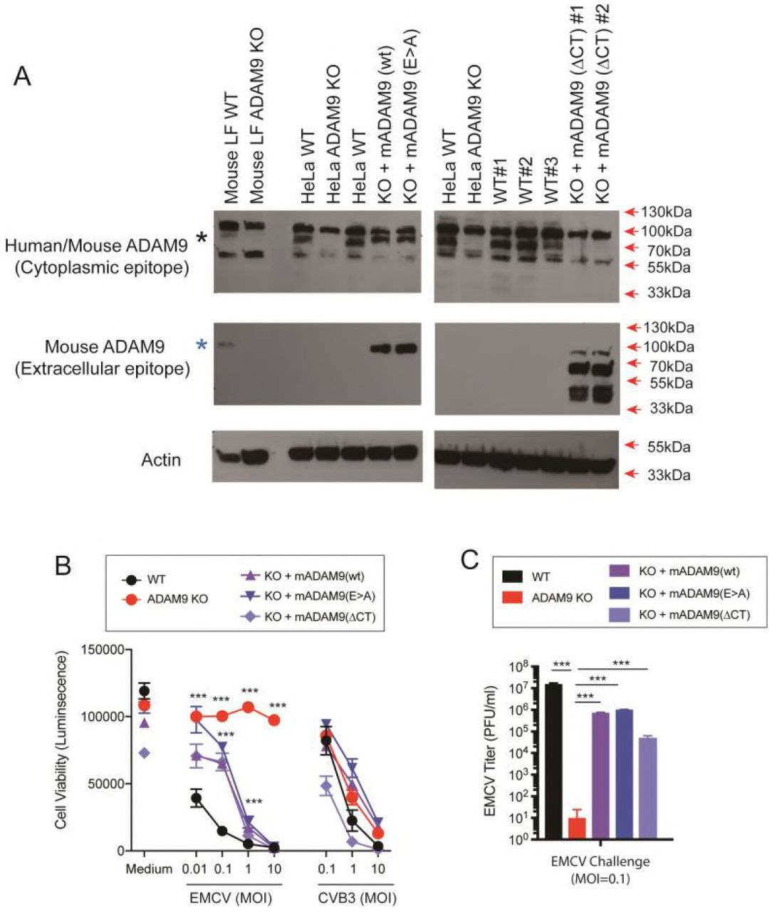 Anti Goat Igg Hrp, supplied by R&D Systems, used in various techniques. Bioz Stars score: 95/100, based on 1 PubMed citations. ZERO BIAS - scores, article reviews, protocol conditions and more https://www.bioz.com/product/rage+function+blocking+antibody/Goat+IgG+HRP-conjugated+Antibody/bio_rxiv__491068-209-4-9 Average 95 stars, based on 1 article reviews
anti goat igg hrp - by Bioz Stars,
2026-10
95/100 stars
|
Buy from Supplier |
|
R&D Systems
human anti vascular cell adhesion molecule vcam 1 Human Anti Vascular Cell Adhesion Molecule Vcam 1, supplied by R&D Systems, used in various techniques. Bioz Stars score: 94/100, based on 1 PubMed citations. ZERO BIAS - scores, article reviews, protocol conditions and more https://www.bioz.com/product/rage+function+blocking+antibody/Human+VCAM-1%2FCD106+Antibody/pm19628926-36-31-37 Average 94 stars, based on 1 article reviews
human anti vascular cell adhesion molecule vcam 1 - by Bioz Stars,
2026-10
94/100 stars
|
Buy from Supplier |
|
R&D Systems
anti laminin 5 antibody Anti Laminin 5 Antibody, supplied by R&D Systems, used in various techniques. Bioz Stars score: 92/100, based on 1 PubMed citations. ZERO BIAS - scores, article reviews, protocol conditions and more https://www.bioz.com/product/rage+function+blocking+antibody/Human+Laminin+alpha+3%2FLaminin-5+Antibody/bio_rxiv__2022__07__28__501930-259-12-15 Average 92 stars, based on 1 article reviews
anti laminin 5 antibody - by Bioz Stars,
2026-10
92/100 stars
|
Buy from Supplier |
|
R&D Systems
isotype matched control goat igg Isotype Matched Control Goat Igg, supplied by R&D Systems, used in various techniques. Bioz Stars score: 96/100, based on 1 PubMed citations. ZERO BIAS - scores, article reviews, protocol conditions and more https://www.bioz.com/product/rage+function+blocking+antibody/Normal+Goat+IgG+Control/pmc05355635__mmc2-273-19-23 Average 96 stars, based on 1 article reviews
isotype matched control goat igg - by Bioz Stars,
2026-10
96/100 stars
|
Buy from Supplier |
|
R&D Systems
anti pd l1 blocking mab Anti Pd L1 Blocking Mab, supplied by R&D Systems, used in various techniques. Bioz Stars score: 93/100, based on 1 PubMed citations. ZERO BIAS - scores, article reviews, protocol conditions and more https://www.bioz.com/product/rage+function+blocking+antibody/Human+PD-L1+Alexa+Fluor%C2%AE+350-conjugated+Antibody/pm39512342-79-29-34 Average 93 stars, based on 1 article reviews
anti pd l1 blocking mab - by Bioz Stars,
2026-10
93/100 stars
|
Buy from Supplier |
|
Becton Dickinson
hamster mabs ha2/5 (function-blocking against mouse β1 integrin Hamster Mabs Ha2/5 (Function Blocking Against Mouse β1 Integrin, supplied by Becton Dickinson, used in various techniques. Bioz Stars score: 90/100, based on 1 PubMed citations. ZERO BIAS - scores, article reviews, protocol conditions and more https://www.bioz.com/product/rage+function+blocking+antibody/cd24+pe+antibody/pmc01415321-61-21-39 Average 90 stars, based on 1 article reviews
hamster mabs ha2/5 (function-blocking against mouse β1 integrin - by Bioz Stars,
2026-10
90/100 stars
|
Buy from Supplier |
Image Search Results
Journal: OncoTargets and Therapy
Article Title:
S100A9 promotes prostate cancer cell invasion by activating TLR4/NF-κB/integrin β1/FAK signaling
doi: 10.2147/ott.s192250
Figure Lengend Snippet: Figure 1 S100A9 promotes prostate cancer cell invasion and β1 integrin expression through interaction with TLR4. PC-3 and DU-145 cells were treated with S100A9 (20 µg/ml) for 48 h. (A) PC-3 and DU-145 cells invasion was measured by transwell invasion assay. (B) The mRNA and protein levels of β1 integrin were determined by qPCR or Western blot. PC-3 and DU-145 cells were treated with S100A9 (20 µg/ml) for 48 h. (C, D) Cell extracts were immunoprecipitated (IP) with control mouse IgG, mouse anti-S100A9 antibody. Immunoblot (IB) was used to detect S100A9, TLR4 and RAGE. PC-3 and DU-145 cells were transfected with TLR4 or control siRNA. (E) TLR4 expression was examined by Western blot after 48 h siRNA transfection. PC-3 and DU-145 cells were transfected with TLR4 or control siRNA followed by stimulation with S100A9. (F) Tumor cell invasion was measured by transwell invasion assay. (G) The mRNA and protein levels of β1 integrin were determined by qPCR or Western blot. Scale bar 50 μ. Magnifcation×200. Data are represented as the mean ± S.E.M. *p<0.05.
Article Snippet: Antibodies and reagents The recombinant
Techniques: Expressing, Transwell Invasion Assay, Western Blot, Immunoprecipitation, Control, Transfection

Journal: OncoTargets and Therapy
Article Title:
S100A9 promotes prostate cancer cell invasion by activating TLR4/NF-κB/integrin β1/FAK signaling
doi: 10.2147/ott.s192250
Figure Lengend Snippet: Figure 2 NF-κB mediates S100A9-induced prostate cancer cell β1 integrin up-regulation. PC-3 and DU-145 cells transfected with or without TLR4 siRNA or control siRNA, were transfected with NF-κB-luciferase reporter plasmid, and treated with S100A9 (20 µg/ml) for 48 h. (A, B) Activity of NF-κB was detected by measuring the relative activity of luciferase. PC-3 and DU-145 cells were treated with or without BAY11-7082 (5µM) for 30 min. Then cells were treated with or without S100A9 (20 µg/ ml) for 48 h. (C) The mRNA and protein levels of β1 integrin were determined by qPCR or Western blot. Data are represented as the mean ± S.E.M. *p<0.05.
Article Snippet: Antibodies and reagents The recombinant
Techniques: Transfection, Control, Luciferase, Plasmid Preparation, Activity Assay, Western Blot

Journal: OncoTargets and Therapy
Article Title:
S100A9 promotes prostate cancer cell invasion by activating TLR4/NF-κB/integrin β1/FAK signaling
doi: 10.2147/ott.s192250
Figure Lengend Snippet: Figure 3 S100A9 promotes prostate cancer cell invasion via integrin β1/FAK signaling. PC-3 and DU-145 cells were treated with or without BAY11-7082 (5µM) for 30 min. Then cells were treated with or without S100A9 (20 µg/ml) for 48 h. (A) Fibronectin expression was determined by Western blot. PC-3 and DU-145 cells were treated with or without S100A9 (20 µg/ml) for 48 h. (B) Supernatant fibronectin (FN) concentration was determined by ELISA. PC-3 and DU-145 cells were treated with or without S100A9 (20 µg/ml) for 30 min. (C) The phosphorylation of FAK was measured by Western blot. PC-3 and DU-145 cells were transfected with control siRNA or integrin β1-specific siRNA for 48 h. Then cells were treated with or without S100A9 (20 µg/ml) for 30 min, the expression of integrin β1 (D) or phosphorylation of FAK (E) was measured by Western blot. PC-3 and DU-145 cells were transfected with control siRNA or integrin β1-specific siRNA for 48 h. Then cells were treated with or without S100A9 (20 µg/ml) for 48 h. (F) The invasion activity were measured by transwell invasion assay. (G) PC-3 and DU- 145 cells were treated with β1 integrin functional blocking antibody MAB13 (50 μg/mL) or control IgG (50 μg/mL) for 30 min and then treated with or without S100A9 (20 µg/ml) for 30 min and the phosphorylation of FAK was measured by Western blot. (H) PC-3 and DU-145 cells were treated with MAB13 (50 μg/mL) or control IgG (50 μg/mL) for 30 min and then treated with or without S100A9 (20 µg/ml) for 48 h for invasion. (I) PC-3 and DU-145 cells were pretreated for 30 min with FAK inhibitor, PF562271 (100 nM) followed by stimulation with S100A9 (20 µg/ml) for 48 h for invasion. Data are represented as the mean ± S.E.M. *p<0.05.
Article Snippet: Antibodies and reagents The recombinant
Techniques: Expressing, Western Blot, Concentration Assay, Enzyme-linked Immunosorbent Assay, Phospho-proteomics, Transfection, Control, Activity Assay, Transwell Invasion Assay, Functional Assay, Blocking Assay

Journal: OncoTargets and Therapy
Article Title:
S100A9 promotes prostate cancer cell invasion by activating TLR4/NF-κB/integrin β1/FAK signaling
doi: 10.2147/ott.s192250
Figure Lengend Snippet: Figure 4 S100A9 induces prostate cancer cell metastasis in vivo. DU-145 cells were transfected with pcDNA3.1 or pc DNA-S100A9 plasmid. (A, B) The expression or secretion of S100A9 was determined by Western blot and ELISA. The cells were injected to nude mice via tail vein. 30 days after inoculation, nude mice were sacrificed. (C) Expressions of S100A9 and integrin β1 in xenograft tumors were detected by immunohistochemistry. (D) Expressions of FAK, p-FAK, NF-kB p65 and p-NF-kB p65 in xenograft tumors were determined by Western blot. (E) Micrometastatic tumors in the lungs of mouse xenografts were counted and subjected to H and E staining. Scale bar 50 μ. Magnifcation×200. Data are represented as the mean ± S.E.M.*p<0.05.
Article Snippet: Antibodies and reagents The recombinant
Techniques: In Vivo, Transfection, Plasmid Preparation, Expressing, Western Blot, Enzyme-linked Immunosorbent Assay, Injection, Immunohistochemistry, Staining
Journal: ACS Omega
Article Title: Hydrogel-Mediated Release of TRPV1 Modulators to Fine Tune Osteoclastogenesis
doi: 10.1021/acsomega.1c06915
Figure Lengend Snippet: Expression and functional analysis of TRPV1 in BMMs and Osteoclasts. (a) BMMs stained for the macrophage marker CD11b (red) and TRPV1 (green) depict the latter’s expression in these cells, both in the absence (upper panel) and presence (lower panel) of RANKL. (b) Expression of TRPV1 (red) in phalloidin-stained osteoclasts (green, upper panel) is confirmed by a peptide segment against anti-TRPV1 antibody (lower panel) that reduces the specific fluorescence signal intensity of the TRPV1 channel.
Article Snippet: For confirming the specificity of the antibody,
Techniques: Expressing, Functional Assay, Staining, Marker, Fluorescence

Journal: ACS Omega
Article Title: Hydrogel-Mediated Release of TRPV1 Modulators to Fine Tune Osteoclastogenesis
doi: 10.1021/acsomega.1c06915
Figure Lengend Snippet: Functional analysis of TRPV1 in BMMs. (a) BMMs were assessed for intracellular Ca 2+ levels upon TRPV1 modulation. Representative intensity profiles of Fluo4-AM intensity at different frames are indicated. (b) Time series graphs of intracellular Fluo4-AM intensities across 200 frames of live imaging. The arrow at the x-axis signifies the time of addition of the respective drugs (20th frame). Gray traces are of individual cells, and the black trace represents the average of 50 cells. (c) Compiled average of different treatments of BMMs, individual cell traces omitted.
Article Snippet: For confirming the specificity of the antibody,
Techniques: Functional Assay, Imaging

Journal: ACS Omega
Article Title: Hydrogel-Mediated Release of TRPV1 Modulators to Fine Tune Osteoclastogenesis
doi: 10.1021/acsomega.1c06915
Figure Lengend Snippet: Functional analysis of TRPV1 in BMMs grown on the CMT:HEMA hydrogel. (a) BMMs grown on hydrogels to check for the endogenous levels of Ca 2+ using Fluo4-AM Ca 2+ -sensitive dye. TRPV1 activation elevates the intracellular Ca 2+ levels, as is quantified in (b); n = 100 cells; one-way ANOVA; ns: non-significant, **** p < 0.0001. (c) Correlation representation of the area of cells and per unit area intensity of Fluo4-AM depicts strong positive correlations under basal and TRPV1-activated conditions but not upon inhibition of the channel.
Article Snippet: For confirming the specificity of the antibody,
Techniques: Functional Assay, Activation Assay, Inhibition

Journal: ACS Omega
Article Title: Hydrogel-Mediated Release of TRPV1 Modulators to Fine Tune Osteoclastogenesis
doi: 10.1021/acsomega.1c06915
Figure Lengend Snippet: Morphological analysis of BMMs grown on the hydrogel. (a) Representative images of BMMs grown on glass or hydrogel in the presence of RANKL and TRPV1 modulators. Right panels denote marked inset of respective images. Phalloidin intensity (b) and morphometric analyses of BMM’s area (c), perimeter (d), length (e), width (f), and LWR (g). n = 18–51 cells per group; one-way ANOVA; ns: non-significant, * p < 0.05, *** p < 0.001, **** p < 0.0001.
Article Snippet: For confirming the specificity of the antibody,
Techniques:

Journal: ACS Omega
Article Title: Hydrogel-Mediated Release of TRPV1 Modulators to Fine Tune Osteoclastogenesis
doi: 10.1021/acsomega.1c06915
Figure Lengend Snippet: Differentiation propensities of BMMs into osteoclasts grown on hydrogel. (a) Representative TRAP assay of BMMs grown on the hydrogel in the presence of the TRPV1 activator (RTX) and inhibitor (5′-IRTX) under differentiating conditions (MCSF + RANKL). (b,c) Quantitation of TRAP-positive cells and multinucleated cells in the presence of capsaicin (b) and RTX (c) shows elevated osteoclastogenesis as compared to MCSF and CMT:HEMA control groups. n = 5–10; one-way ANOVA; ** p < 0.01, *** p < 0.005, **** p < 0.001.
Article Snippet: For confirming the specificity of the antibody,
Techniques: TRAP Assay, Quantitation Assay
Journal:
Article Title: Sequence-Specific Interaction between the Disintegrin Domain of Mouse ADAM 3 and Murine Eggs: Role of ?1 Integrin-associated Proteins CD9, CD81, and CD98
doi:
Figure Lengend Snippet: Binding of beads coated with mADAM 3 disintegrin domains: effects of anti-integrin subunit antibodies. Fluorescent beads were coated with, as indicated, mADAM 3 or hADAM 15 disintegrin domains made in either E. coli (E-ADAM) or Drosophila cells (D-ADAM) as described in the legend to Figure Figure3.3. Eggs were preincubated for 30 min at 37°C with, as indicated, either: buffer (A1, A4, and A7); function blocking anti α6 mAb, GoH3 (A2, A5, and A8); nonfunction blocking anti-α6 mAb, J1B5 (A3, A6, and A9); hamster IgG (B1 and B4); hamster anti-mouse β3 mAb (B2 and B5), or hamster anti-mouse αv mAb (B3 and B6) at 200 μg/ml, with the exception that the eggs for Drosophila ADAM3 bead binding were treated with GoH3 at 100 μg/ml. Good inhibition of binding of beads coated with Drosophila ADAM 3 was also observed with 50 μg/ml GoH3. Bead binding was then observed in a fluorescence microscope as described in the legend to Figure Figure3.3. Relative fluorescence intensities, normalized to the buffer control for each set (A1, A4, A7, B1, B4), were determined as described in MATERIALS AND METHODS and then plotted (to the right of the micrographs); numbers under each bar refer to the respective micrograph panels.
Article Snippet: The function-blocking
Techniques: Binding Assay, Blocking Assay, Inhibition, Fluorescence, Microscopy

Journal:
Article Title: Sequence-Specific Interaction between the Disintegrin Domain of Mouse ADAM 3 and Murine Eggs: Role of ?1 Integrin-associated Proteins CD9, CD81, and CD98
doi:
Figure Lengend Snippet: Effects of antibodies to β1 integrin-associated proteins on fertilization and ADAM 3 bead binding. (A) Zona-free eggs were processed for immunofluorescence with mAbs against CD81 and CD98 as described in MATERIALS AND METHODS. The control antibody for CD81 was hamster IgG and that for CD98 was rat IgG. Sperm-egg binding (B) and fusion (C) were assayed following preincubation with 200 μg/ml the indicated antibodies as described in the legend to Figure Figure2.2. The data in B and C are from one experiment. The experiment was repeated three times with similar results. (D) Fluorescent beads coated with E. coli mADAM 3 disintegrin domain were processed for binding to zona-free eggs following a 30-min preincubation with 200 μg/ml the indicated antibodies as described in MATERIALS AND METHODS. The sample labeled control was preincubated with buffer. Scion image analysis (graph to the right in D) was performed as described in the legend to Figure Figure6. 6. The anti-CD81 antibody used in the experiments shown was the mAb 2F7.
Article Snippet: The function-blocking
Techniques: Binding Assay, Immunofluorescence, Labeling
Journal: Cell reports
Article Title: Neurokinin-1 Receptor Signaling Is Required for Efficient Ca 2+ Flux in T-Cell-Receptor-Activated T Cells
doi: 10.1016/j.celrep.2020.02.054
Figure Lengend Snippet: KEY RESOURCES TABLE
Article Snippet:
Techniques: Functional Assay, Recombinant, Staining, Avidin-Biotin Assay, Blocking Assay, Plasmid Preparation, Inhibition, Fluorescence, SYBR Green Assay, Activation Assay, Bicinchoninic Acid Protein Assay, In Situ, Enzyme-linked Immunosorbent Assay, Activity Assay, Labeling, Software
Journal: Reproduction, Fertility and Development
Article Title: The involvement of hypoxia-inducible factor 1α (HIF1α)-stabilising factors in steroidogenic acute regulatory (STAR) protein-dependent steroidogenesis in murine KK1 granulosa cells
doi: 10.1071/rd21170
Figure Lengend Snippet: Fig. 1. Effects of hypoxia and functional suppression of HIF1-complexes on HIF1α and STAR expression in KK1 granulosa cells. Cells were stimulated with 0.1 mM dbcAMP and treated with 5 nM echinomycin (Echino/E, functional blocker of HIF1-complexes) under 20% and 1% O2. HIF1α (a) (115 kDa) and STAR (b) (30 kDa) protein expression as determined by western blot analysis. The results were normalised against ACTB and calculated as the average SOD. Representative western blots are shown. For (a) and (b) one- way ANOVA was applied (P < 0.0001 each), followed by a Tukey–Kramer multiple com parisons post-test. Bars with asterisks differ at: *P < 0.05, **P < 0.01, ***P < 0.001 as indicated on the figure. P < 0.05 was considered significant. Results are presented as mean ± s.d.
Article Snippet: Antigen Product no. Producer Dilution Species/type STAR Dr. DM Stocco, Texas Tech University Health Science Center, 1:5000 Rabbit, polyclonal Lubbock, TX, USA Clark et al. (1994)
Techniques: Functional Assay, Expressing, Western Blot

Journal: Reproduction, Fertility and Development
Article Title: The involvement of hypoxia-inducible factor 1α (HIF1α)-stabilising factors in steroidogenic acute regulatory (STAR) protein-dependent steroidogenesis in murine KK1 granulosa cells
doi: 10.1071/rd21170
Figure Lengend Snippet: Fig. 2. Basal mRNA and protein expression of HIF1α-stabilising factors: FIH (42 kDa), VHL (17 kDa), PHD1 (44 kDa), PHD2 (50 kDa) and PHD3 (27 kDa) in murine ovaries, KK1 granulosa cells and COS cells serving as positive controls. Representative western blots are shown. (a) Results from qualitative PCR. (b) Protein expression determined by western blot. ACTB was used as loading control.
Article Snippet: Antigen Product no. Producer Dilution Species/type STAR Dr. DM Stocco, Texas Tech University Health Science Center, 1:5000 Rabbit, polyclonal Lubbock, TX, USA Clark et al. (1994)
Techniques: Expressing, Western Blot, Control

Journal: Reproduction, Fertility and Development
Article Title: The involvement of hypoxia-inducible factor 1α (HIF1α)-stabilising factors in steroidogenic acute regulatory (STAR) protein-dependent steroidogenesis in murine KK1 granulosa cells
doi: 10.1071/rd21170
Figure Lengend Snippet: Fig. 4. Effects of changing oxygen content (20%, 10%, 1% O2) and functional blocking of HIF1α on FIH and VHL in KK1 granulosa cells. Cells were stimulated with 0.3 mM dbcAMP and treated with echinomycin (Echino/E). Western blot analysis was performed. COS cells served as positive controls. Representative immunoblots are shown. Expression of FIH (a) and VHL (b) were examined, normalised against ACTB and calculated as the average SOD. One-way ANOVA was applied: FIH (P = 0.92), VHL (P = 0.96). P < 0.05 was considered significant. Results are presented as mean ± s.d.
Article Snippet: Antigen Product no. Producer Dilution Species/type STAR Dr. DM Stocco, Texas Tech University Health Science Center, 1:5000 Rabbit, polyclonal Lubbock, TX, USA Clark et al. (1994)
Techniques: Functional Assay, Blocking Assay, Western Blot, Expressing

Journal: Reproduction, Fertility and Development
Article Title: The involvement of hypoxia-inducible factor 1α (HIF1α)-stabilising factors in steroidogenic acute regulatory (STAR) protein-dependent steroidogenesis in murine KK1 granulosa cells
doi: 10.1071/rd21170
Figure Lengend Snippet: Fig. 5. Effects of changing oxygen content (20%, 10%, 1% O2) and functional blocking of HIF1α on PHD1, PHD2 and PHD3 in KK1 granulosa cells. Cells were stimulated with 0.3 mM dbcAMP and treated with echinomycin (Echino/E). Western blot analysis was performed. COS cells served as positive controls. Representative immunoblots are shown. Expression of PHD1 (a), PHD2 (b) and P HD3 (c) was examined, normalised against ACTB and calculated as the average SOD. (a, b) One- way ANOVA was applied revealing P = 0.8 for PHD1 (a), and P < 0.0001 for PHD2 (b), in case of P < 0.05, followed by a Tukey–Kramer multiple comparisons post-test: *P < 0.001. Results are presented as mean ± s.d.
Article Snippet: Antigen Product no. Producer Dilution Species/type STAR Dr. DM Stocco, Texas Tech University Health Science Center, 1:5000 Rabbit, polyclonal Lubbock, TX, USA Clark et al. (1994)
Techniques: Functional Assay, Blocking Assay, Western Blot, Expressing

Journal: Reproduction, Fertility and Development
Article Title: The involvement of hypoxia-inducible factor 1α (HIF1α)-stabilising factors in steroidogenic acute regulatory (STAR) protein-dependent steroidogenesis in murine KK1 granulosa cells
doi: 10.1071/rd21170
Figure Lengend Snippet: Fig. 6. The effect of functional blocking of PHDs on HIF1α and STAR protein expression in KK1 granulosa cells. Cells were stimulated with 0.3 mM dbcAMP and PHD function was blocked using increasing dosages of roxadustat (25 μM, 50 μM and 100 μM); HIF1α activity was blocked with echinomycin (Echino). HIF1α (a) and STAR (b) protein levels were examined by western blot. Representative immunoblots are shown. The protein expression was normalised against ACTB and calculated as the average SOD. One-way ANOVA was applied (P < 0.0001 each), followed by a Tukey–Kramer multiple comparisons post-test. P < 0.05 was considered significant. Results are presented as mean ± s.d. Bars with asterisks differ at: *P < 0.05, **P < 0.01, ***P < 0.001 as indicated on the figure.
Article Snippet: Antigen Product no. Producer Dilution Species/type STAR Dr. DM Stocco, Texas Tech University Health Science Center, 1:5000 Rabbit, polyclonal Lubbock, TX, USA Clark et al. (1994)
Techniques: Functional Assay, Blocking Assay, Expressing, Activity Assay, Western Blot

Journal: Reproduction, Fertility and Development
Article Title: The involvement of hypoxia-inducible factor 1α (HIF1α)-stabilising factors in steroidogenic acute regulatory (STAR) protein-dependent steroidogenesis in murine KK1 granulosa cells
doi: 10.1071/rd21170
Figure Lengend Snippet: Fig. 7. Schematic representation of proposed regulation of HIF1α availability and its importance in STAR-dependent steroidogenesis in granulosa cells. HIF1α expression and the activity of HIF1-complexes, are requested for STAR expression and steroidogenesis. VHL, FIH and PHDs, modulate (inhibit) the presence of HIF1α depending on the availability of O2. However, FIH and VHL are expressed in an O2-independent manner in granulosa cells, while PHD2 (but not PHD1 or PHD3) responds to changing O2 levels by increasing its expression. Enhanced PHD2 activity under ambient and/or moderately lowered O2 (20% and 10% O2) appears to have a protective role in regulating HIF1α availability by preventing an exaggeratedly high expression. When overexpressed, e.g. during severely lowered (1%) O2, HIF1α exerts negative effects over STAR expression and steroidogenesis. With this, the effects of HIF1α appear to be biphasic, i.e. while it is required for STAR expression, when overexpressed it exerts negative effects. The underlying molecular mechanisms remain to be elucidated.
Article Snippet: Antigen Product no. Producer Dilution Species/type STAR Dr. DM Stocco, Texas Tech University Health Science Center, 1:5000 Rabbit, polyclonal Lubbock, TX, USA Clark et al. (1994)
Techniques: Expressing, Activity Assay
Journal: bioRxiv
Article Title: Identification of A Disintegrin and Metalloproteinase 9 domain (ADAM9) required in the early stages of encephalomyocarditis virus infection
doi: 10.1101/491068
Figure Lengend Snippet: WT and ADAM9 KO HeLa cells were transduced with retroviral vectors with wild-type (WT) murine ADAM9 (mADAM9), catalytically inactive mutant ADAM9 (E>A), cytoplasmic tail deleted (ΔCT) ADAM9 constructs, or GFP control vectors. Transduced cells were selected and cloned by limiting dilution, and expression of mADAM9 was confirmed by western blot analysis. (A) WT, KO and rescue cell lysates were prepared in RIPA buffer and run on 12% SDS-PAGE gels and transferred to PVDF membranes. Membranes were blocked with 3% BSA and incubated with ADAM9 antibodies. Upper panel, rabbit anti-human ADAM9 (Cell signaling #2099) that detects an epitope in the intracellular domain of human ADAM9 and cross-reacts with mouse ADAM9. Middle panel, goat anti-mouse ADAM9 (R&D systems AF949) that detects the extracellular domain of murine ADAM9, but not human ADAM9. Lower panel, anti-Actin (Santa Cruz sc-1616) which detects both human and murine ϟ-Actin. Bands were visualized using HRP and ECL reagent. Upper panel: WT but not KO cells expressed human ADAM9. Middle panel: Rescue but not KO cells expressed murine ADAM9. Lower panel: Actin loading control. (B) WT clones, ADAM9 KO clones and rescued ADAM9-expressing clones were infected with EMCV or CVB3 at varying MOI and incubated at 37 °C for 24 h. Viability of EMCV-infected and CVB3-infected clones was measured by CellGlo ATP luminescence. (C) EMCV replication was quantified in infected culture supernatants by plaque assay using BHK-21 cells. Neither the functional sequence of the ADAM9 metalloproteinase domain nor the cytoplasmic tail are required for EMCV infection. ***, P <0.0001, KO vs. WT; KO vs. rescue.
Article Snippet: 3% BSA blocking buffer),
Techniques: Transduction, Mutagenesis, Construct, Clone Assay, Expressing, Western Blot, SDS Page, Incubation, Infection, Plaque Assay, Functional Assay, Sequencing